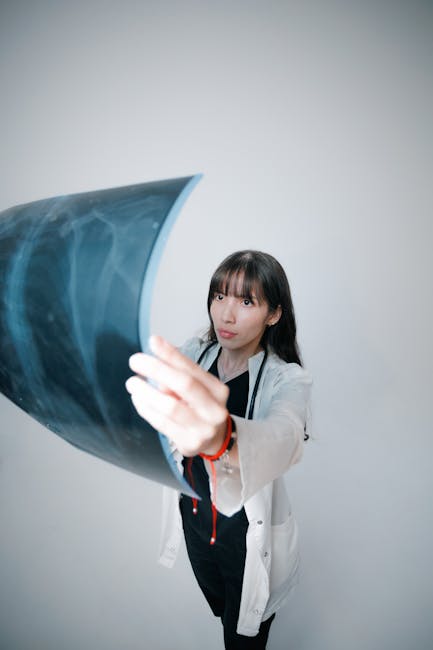
Revolution in Radiology: AI’s Journey from Lab to Clinic
Grab a cup of your favorite brew and let’s explore how a powerful technology has been transforming radiology, one pixel at a time. It’s a story of groundbreaking research, significant advancements, and a fundamental shift in how medical professionals approach diagnostics and patient care.
The Dawn of AI in Radiology (2010s)
The early 2010s marked the genesis of a revolution. The first stirrings of this new technology were beginning to ripple through the halls of academia and research institutions. Eager scientists were crafting algorithms that could learn from vast amounts of data—images, primarily—and identify patterns invisible to the naked eye. This nascent stage was characterized by experimentation and a burgeoning sense of possibility.
One of the earliest adopters was Dr. Matthew Lungren at Stanford University. He started tinkering with this technology in radiology around 2012, initially using it to classify lung nodules on CT scans. Little did he know that this would be just the first step into a fascinating new world – a world poised to fundamentally change medical imaging.
The Breakthrough (Mid-2010s)
As we moved into the mid-2010s, momentum began to build. This period witnessed a surge in research and development, with algorithms becoming more accurate, efficient, and reliable. A key development during this time was the rise of deep learning, a subset of machine learning that mimics how humans learn, and its subsequent application to medical imaging.
In 2015, Dr. Stephen Solomon’s team at NYU Langone Medical Center published a landmark study using this technology to detect pneumothorax (collapsed lung) on chest X-rays. This wasn’t just about improving diagnostics; it was about saving lives by detecting potentially fatal conditions faster and more accurately – a testament to the potential of this evolving field.
AI Meets the Clinic (Late 2010s – Early 2020s)
The late 2010s and early 2020s represented a pivotal shift, as research moved beyond the laboratory and began to impact clinical practice. Hospitals started integrating systems powered by these algorithms into their workflows, leading to improvements in both patient care and workflow efficiency. This period was defined by real-world applications and growing acceptance within the medical community.
- 2018: Google’s DeepMind created an algorithm capable of interpreting optical coherence tomography (OCT) scans – a crucial tool in managing eye diseases like glaucoma – with a speed and accuracy on par with experienced specialists.
- 2019: Arterys, a California-based company, gained FDA approval for its algorithm-powered cardiac MRI analysis system. This technology can analyze heart scans in seconds, freeing up radiologists’ time to focus on other tasks and enhancing overall productivity.
AI Today: A Radiologist’s New Best Friend
Today, the technology is an integral part of radiology departments worldwide. It’s helping radiologists work more efficiently, make more accurate diagnoses, and provide better patient care. This integration has led to a more collaborative approach, where human expertise and algorithmic precision work in tandem to achieve optimal patient outcomes.
- Faster Reporting: Algorithms can preprocess images, prioritize cases based on urgency, and even draft reports, enabling radiologists to read more exams in less time.
- Improved Diagnostics: By learning from vast amounts of data, these systems can detect subtle abnormalities that humans might miss. It’s like having a second set of eyes – one with enhanced capabilities.
- Personalized Medicine: This technology is helping tailor treatments to individual patients by providing insights into disease progression and response to therapy, paving the way for more targeted and effective medical interventions.
The Future: AI in Radiology
Looking ahead, the possibilities are truly remarkable. Imagine a future where these systems assist radiologists in real-time during procedures like MRI or CT scans, providing immediate feedback and guidance. The potential extends to AI-driven tools for 3D reconstruction and virtual reality navigation during interventions, making complex procedures more accessible and precise.
Furthermore, algorithms that can predict patient outcomes and guide clinicians towards the most appropriate treatment plans are on the horizon, promising a new era of proactive and personalized healthcare. This is the journey of how this technology has evolved, from its humble beginnings in research labs to its current status as a radiologist’s trusted ally. It’s an exciting time for both the technology and medicine, isn’t it? Here’s to embracing change and making healthcare better, one pixel at a time!


Leave a Reply
You must be logged in to post a comment.